
By: Robert L. Bard, MD / Edited by: Lennard M. Goetze, Ed.D & Roberta Kline, MD
Introduction:
Male breast cancer remains one of the most under-recognized and
under-diagnosed cancers, often dismissed due to societal bias or lack of
awareness. However, advancements in imaging technologies are revolutionizing
early detection, staging, and treatment monitoring. At the forefront of this
transformation is the integration of high-resolution ultrasound and
Doppler-based vascular imaging—a non-invasive, real-time solution offering
critical insight into tumor biology, particularly in high-risk male populations
such as first responders and those exposed to occupational toxins.
While mammography remains a conventional screening tool, it is rarely used in men unless they present with symptoms such as breast enlargement, nipple discharge, or palpable masses—particularly in those with a BRCA gene mutation. More often, practitioners now rely on ultrasound as the primary imaging modality for male breast evaluation. This includes the use of advanced Doppler to assess vascular activity, which can help distinguish benign growths from malignancies.
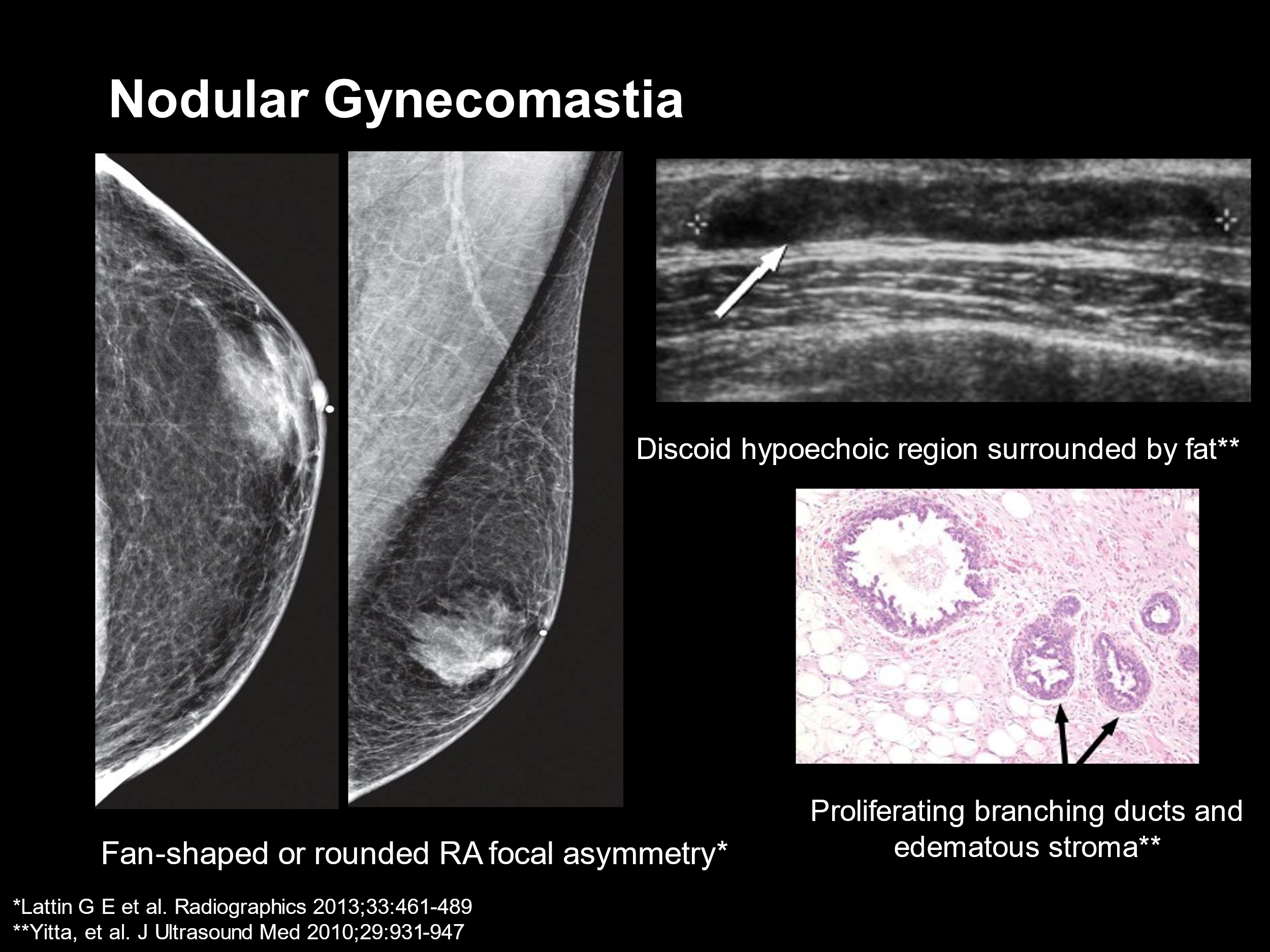
Common benign findings such as
gynecomastia—especially in adolescent and young adult males—are readily identified
with ultrasound. Fatty tumors like lipomas also appear clearly using this
modality. However, the diagnostic power of ultrasound becomes critical when
suspicious symptoms present, such as subareolar masses, nipple retraction, or
bloody discharge. In such cases, ultrasound can quickly detect the presence,
size, and structure of the tumor and guide further intervention.
The Role of Vascular Imaging
Color Doppler ultrasound enhances the ability to characterize lesions by
mapping their blood flow. Malignant tumors often display increased vascularity,
with irregular, tortuous vessels visible within or around the lesion. This
vascular signature, known as neovascularity, has been recognized since the
1990s as a biomarker for tumor aggression.
In clinical practice, once a suspicious lesion
is visualized, additional imaging such as MRI or PET-CT may be employed.
However, Doppler ultrasound allows for immediate evaluation without radiation
exposure. Notably, it enables the patient to point to areas of pain or concern,
allowing for focused and accurate real-time scanning.

Not all tumors behave predictably. Some papillary carcinomas can degenerate into cystic masses. Others may swell during treatment due to inflammation, edema, or immune system response—even as they are responding to therapy. Understanding these patterns is vital to avoid misinterpreting progression when, in fact, healing is occurring.
When used longitudinally, Doppler ultrasound
enables clinicians to monitor the regression of tumor vasculature during
treatment. This provides a quantitative assessment—measuring tumor vessel
density in percentages—that can guide whether therapy is effective or needs to
be adjusted. For instance, a patient may present with a vascular index of 4.5%
pre-treatment, which may drop to 1% after several months of targeted therapy.
This objective measurement offers far more accuracy than visual inspection or
palpation alone.
Minimally Invasive Treatment Monitoring
Several non-surgical treatment options rely heavily on imaging guidance
for planning and efficacy evaluation. Technologies such as High-Intensity
Focused Ultrasound (HIFU), cryoablation, and radiofrequency ablation (RFA) use
heat or cold to destroy tumor tissue. In these cases, vascular imaging confirms
the procedure’s success by showing an immediate absence of tumor blood flow.
Other slower-acting treatments—such as
antioxidants, hormone therapy, or radiation—may take weeks or months to show
regression. In such instances, continued ultrasound surveillance is critical.
Blood flow patterns are monitored over time to determine whether the lesion is
resolving, stabilizing, or growing.

Ultrasound's versatility extends beyond cancer detection. Post-surgical complications such as lymphoceles, infections, or scar tissue entrapment can also be identified. Chronic pain after breast surgery in men is often due to nerve entrapment within scar tissue—a diagnosis more readily made through high-resolution ultrasound than MRI. Similarly, benign findings such as calcified scars or foreign bodies (e.g., splinters or surgical remnants) may mimic malignancy but can be quickly differentiated with imaging.
Lymphomas and other rare cancers may also
appear in the male breast or axillary region. These can be identified with the
same sonographic techniques, providing a complete diagnostic pathway from
primary evaluation to follow-up and surveillance.

A key benefit of blood flow-guided imaging is the ability to target biopsies more precisely. By identifying areas of active tumor vascularity, clinicians can avoid sampling necrotic or non-diagnostic tissue. This not only improves diagnostic yield but also reduces the risk of complications. In one example, a soft, movable lump was initially presumed to be a benign fatty tumor. However, Doppler imaging revealed a rich vascular supply and proximity to the aorta—making blind biopsy potentially dangerous and underscoring the life-saving utility of vascular guidance.
Conclusion: Transforming the Paradigm of
Male Breast Cancer
Modern ultrasound technologies, particularly when paired with vascular
flow analytics, have elevated male breast cancer diagnostics from a reactive,
often delayed process to one that is fast, precise, and patient-centered. From
detecting suspicious lumps to guiding therapies and tracking progress, these
tools empower clinicians with real-time data to personalize care and avoid
unnecessary procedures.
In a population that has long been
underrepresented in breast cancer screening campaigns, the integration of
advanced imaging offers not only a new level of clinical insight but also a
renewed hope for earlier detection and better outcomes. As the field continues
to evolve, image-guided care may well become the gold standard for managing
male breast cancer and its many clinical challenges.


No comments:
Post a Comment